Innovative imaging research in rheumatology
Cutting-edge approaches and strategies
Department of Rheumatology und Clinical Immunology (Clinic of Intern. Med. III), University Hospital Bonn
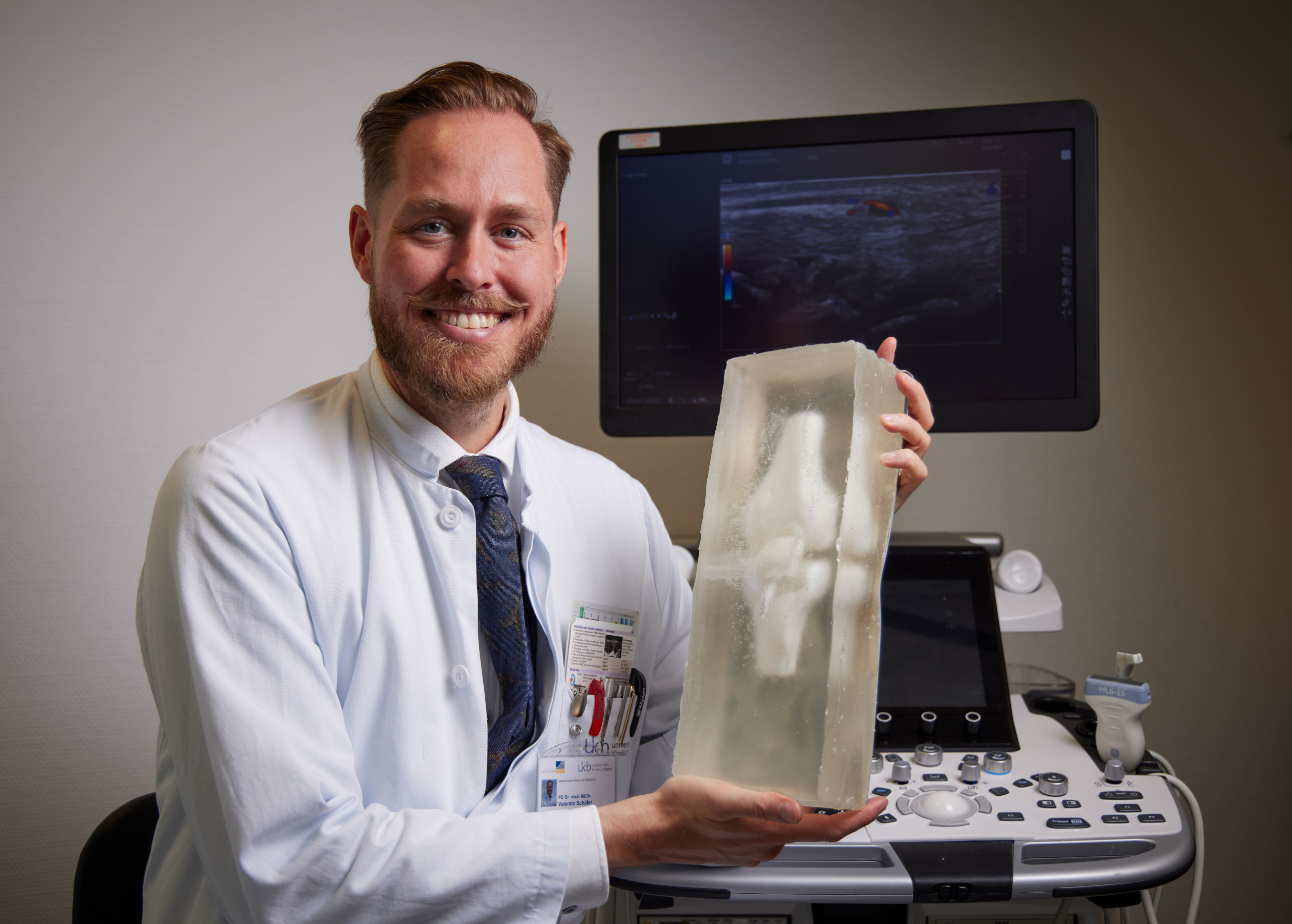
Prof. Dr. MUDr. Valentin S. Schäfer
Prof. Valentin Schäfer and his research group members perform a broad spectrum of highly innovative imaging research in the field of rheumatologic inflammatory processes and beyond.
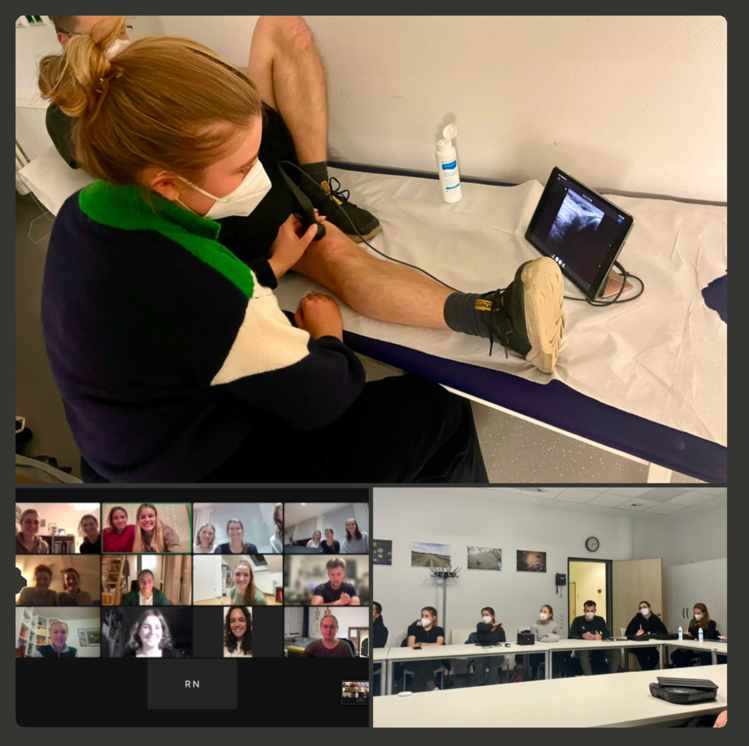
Particular focus is on ultrasound research: This includes investigations of state-of-the-art portable ultrasound devices that employ a novel handheld probe technology for point-of-care ultrasound (POCUS), i.e. location-unbound ultrasound diagnostics, but includes also the development of ultrasound training models (for a realistic reproduction of joints and vessels in healthy and pathologically altered states) using the latest 3D printing technology, and the combination of both tools to break new ground in medical student education and telemedical training of practical ultrasound skills, as for example within the TELUS and TELMUS studies.
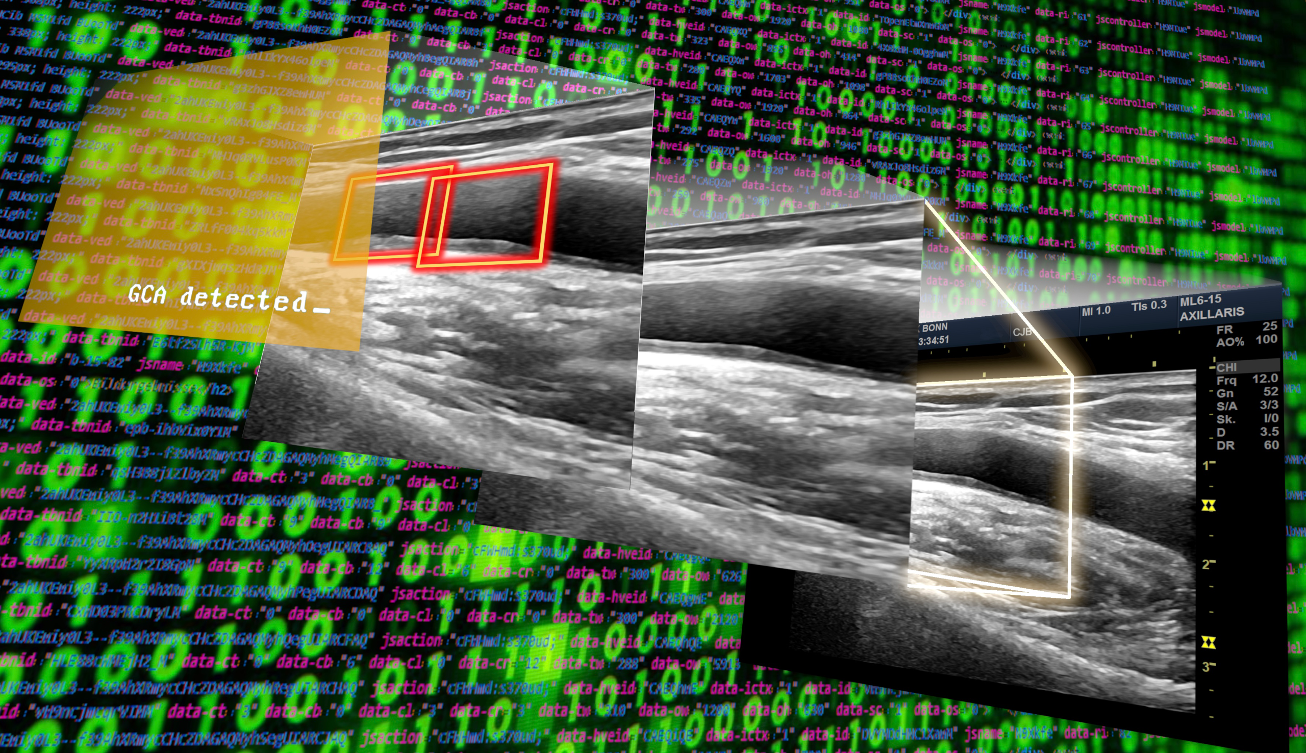
Furthermore, in collaboration with the Albarqouni-Lab, the team around PD Dr. Valentin Schäfer and Claus-Jürgen Bauer is working on an AI assistance in the early ultrasound detection of rheumatological emergencies.
Beyond ultrasound research, a variety of imaging approaches are being investigated, for example dual energy computed tomography for gout diagnosis, or improved PET-CT imaging of vascular inflammation activity utilizing novel VAP-1 tracers. In large research projects on giant cell arteritis, polymyalgia rheumatica, rheumatoid and psoriatic arthritis, structured multimodal imaging (at different stages of disease progression) is linked with genetic and immunological biomarker analyses as well as synovial biopsy investigations of cellular and non-cellular processes at the site of active joint inflammation.
Methods
- Sonography: focus on ultrasound of vessels and joints
- Sonography: focus on (contrast-enhanced) vascular ultrasound of the aorta and its branches.
- MRI: focus on aortic/ cardiac and cerebral imaging
- DECT (Dual Energy Computed Tomography)
- Ultrasound models and 3D printing
- Ultrasound Scoring System (SOLAR Score)
- State-of-the-art Point of Care Ultrasound-device application
- Quantitative analysis of a new fluorescence imaging ( Rheuma Scan)
- Neural network development (Artificial Intelligence)
- Teledidactics
- Synovial biopsy
- Flow cytometry for identification of cellular infiltrates
- ELISA for the determination of cytokine signaling pathways
- Transcriptome analyses